TEE

Transesophageal Echocardiography (TEE) Simulation Training


Course Image
Course Overview

The Transesophageal Echocardiography Simulation Training Program is designed to demystify TEE by focusing on probe control, spatial orientation, and structured image acquisition without patient risk. The course addresses one of the steepest learning curves in echocardiography-understanding where the probe is and how to obtain the desired view safely.

Through immersive simulation, participants learn standard TEE views, cardiac anatomy relationships, and Doppler applications using real patient datasets. The program emphasizes safety, efficiency, and diagnostic accuracy, preparing learners for perioperative, critical care, and cardiac settings.

Simulation-based repetition allows participants to convert complex probe maneuvers into controlled, purposeful movements- building competence before performing TEE on live patients.

Learning Objectives
  • Understand indications, limitations, and safety principles of TEE
  • Develop safe and controlled TEE probe manipulation techniques
  • Recognize cardiac anatomy through standard transesophageal views
  • Acquire systematic mid-esophageal and transgastric imaging planes
  • Apply Doppler techniques for transesophageal hemodynamic assessment
  • Identify common intra-cardiac and valvular pathologies
  • Organize TEE findings into structured diagnostic sequences
Learning Methodology
  • High-fidelity TEE simulator with real patient datasets
  • Stepwise probe navigation and view acquisition training
  • Faculty-supervised hands-on simulation sessions
  • 3D anatomical visualization linked to probe movement
  • Case-based pathology interpretation
  • Performance analytics and image review
Learning Outcomes
  • Perform a complete standard TEE examination in a simulated environment
  • Maintain consistent image quality across standard TEE views
  • Demonstrate safe probe handling without simulated injury events
  • Accurately recognize and report common TEE pathologies
  • Communicate TEE findings clearly in perioperative and ICU settings
  • Enter supervised clinical TEE practice with improved confidence
Eligibility Criteria
  • Cardiac anesthesiologists and anesthesia trainees
  • Critical care and perioperative physicians
  • Cardiology fellows
  • Echocardiography trainees
  • Clinicians involved in perioperative or ICU cardiac assessment
Course Modules
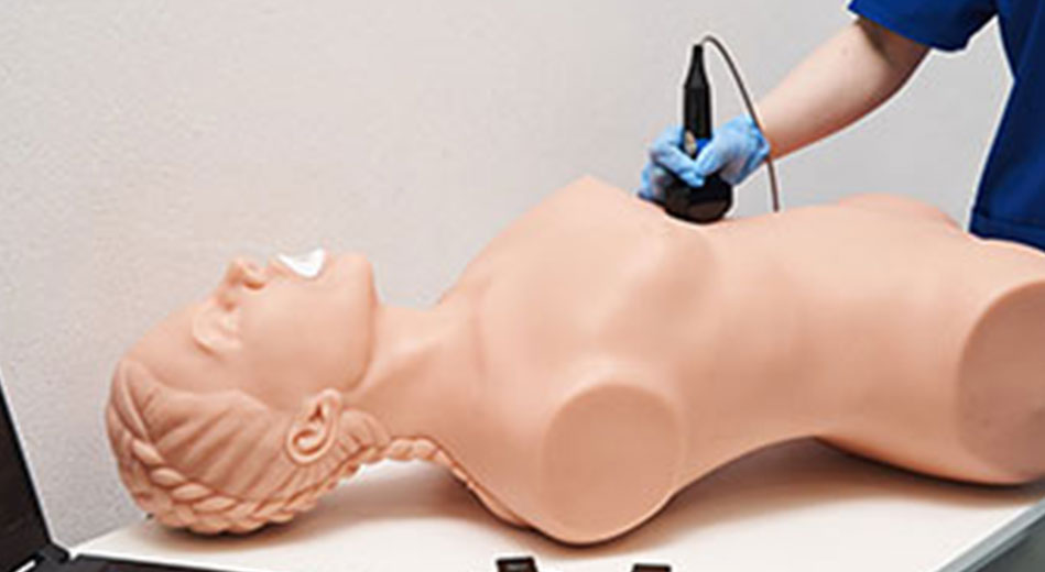
Module 1: TEE Fundamentals & Safety
  • Principles of TEE ultrasound physics
  • Indications and contraindications for TEE
  • Patient safety, probe handling, and ergonomics
  • Infection control and procedural precautions
Module 2: TEE Probe Handling & Anatomical Orientation
  • Introduction to TEE probe controls and movements
  • Depth, flexion, rotation, and omniplane manipulation
  • 3D cardiac anatomy correlation with TEE views
  • Real-time probe tracking in simulation environment
Module 3: Standard TEE Views & Image Acquisition
  • Mid-esophageal views
  • Transgastric views
  • Upper esophageal views
  • Valve anatomy and spatial orientation
  • Stepwise acquisition of standard TEE windows
  • Pre-recorded real patient TEE datasets
Module 4: Doppler Applications in TEE
  • Color Doppler for valve assessment
  • Pulse Wave Doppler (PWD) and Continuous Wave Doppler (CWD)
  • Flow velocity analysis and interpretation
  • Identification of valvular and shunt pathologies
Module 5: Advanced TEE Functional & Hemodynamic Assessment
  • Left and right ventricular functional evaluation via TEE
  • Valve morphology and pathology assessment
  • Perioperative and peri-arrest cardiac assessment
  • Focused TEE protocols (FoCUS-TEE concepts)
Module 6: Clinical Decision-Making & Reporting
  • Case-based TEE interpretation (Normal & Abnormal)
  • Structured TEE reporting
  • Measurement documentation and image archiving
  • Performance analytics and competency assessment

Quick Contact

WhatsApp