Emergency Ultrasound (e-FAST)

  • Home
  • Emergency Ultrasound (e-FAST)

Emergency Ultrasound (e-FAST) Simulation Training Program


Course Image
Course Overview

The Emergency Ultrasound (e-FAST) Simulation Training Program is designed to train clinicians in rapid, structured ultrasound assessment for trauma and acute care scenarios. The course focuses on developing speed, accuracy, and decision-oriented scanning using the extended Focused Assessment with Sonography for Trauma (e-FAST) protocol.

Through high-fidelity simulation, participants learn to detect free fluid in abdominal, pelvic, and thoracic spaces while maintaining strict protocol adherence under time pressure. Realistic trauma scenarios enable learners to practice systematic scanning, image interpretation, and prioritization without patient risk.

This program prepares clinicians to integrate e-FAST confidently into emergency workflows, supporting timely clinical decisions in high-stakes environments.

Learning Objectives
  • Understand the principles and clinical role of the e-FAST protocol
  • Develop rapid transducer placement and probe orientation skills
  • Identify key anatomical windows used in trauma ultrasound
  • Apply standardized e-FAST scanning sequences
  • Differentiate normal findings from free fluid and pleural pathology
  • Correlate ultrasound findings with trauma mechanisms
  • Perform protocol-driven ultrasound assessments under simulated time constraints
Learning Methodology
  • High-fidelity emergency ultrasound simulator–based training
  • Faculty-supervised hands-on scanning sessions
  • Protocol-based stepwise skill development
  • Real-time B-mode imaging with trauma case simulation
  • Combined abdominal, pelvic, and thoracic scenario practice
  • Objective performance feedback and case review
Learning Outcomes
  • Perform a complete e-FAST examination accurately in a simulated emergency setting
  • Maintain protocol consistency across multiple trauma scenarios
  • Detect and localize free fluid and pleural effusions with confidence
  • Demonstrate improved scanning speed without loss of image quality
  • Communicate e-FAST findings clearly during trauma case review
  • Enter supervised emergency ultrasound practice with enhanced situational readiness
Eligibility Criteria
  • Emergency medicine physicians and residents
  • Trauma surgeons and surgical trainees
  • Critical care and intensive care clinicians
  • Anesthesiologists involved in trauma care
  • Radiology trainees
  • Medical officers involved in emergency and trauma response
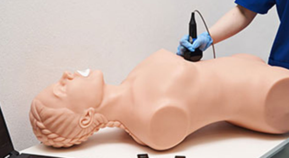
Course Modules
Module 1: e-FAST Fundamentals
  • Indications and scope of e-FAST
  • Patient positioning and probe selection
  • Image orientation and scanning ergonomics
Module 2: Abdominal Free Fluid Assessment
  • Morrison’s pouch evaluation
  • Perihepatic space assessment
  • Identification of minimal and large volume free fluid
Module 3: Splenic & Pelvic Windows
  • Splenorenal recess and perisplenic space evaluation
  • Pelvic cavity scanning
  • Differentiation of minimal versus significant fluid collections
Module 4: Thoracic & Pleural Evaluation
  • Left and right pleural scanning
  • Detection of pleural effusions
  • Integration of thoracic findings into the e-FAST protocol
Module 5: Integrated Trauma Scenarios
  • Combined abdominal and pelvic free fluid cases
  • Abdominal free fluid with pleural effusion
  • Bilateral pleural effusion scenarios
  • Protocol completion under simulated emergency conditions

Quick Contact

WhatsApp